Facilities
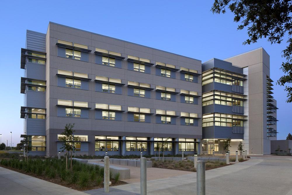
We are located in VM3B on the School of Veterinary Medicine on the UC Davis campus and consists of an open laboratory collaborative model with specialized rooms for embryo and sperm microscopy and cell culture as well as an open space with more than 10 laboratory bench stations, a liquid nitrogen storage and programmable cell freezer, as well as two biosafety cabinets and laminar-flow workstations.
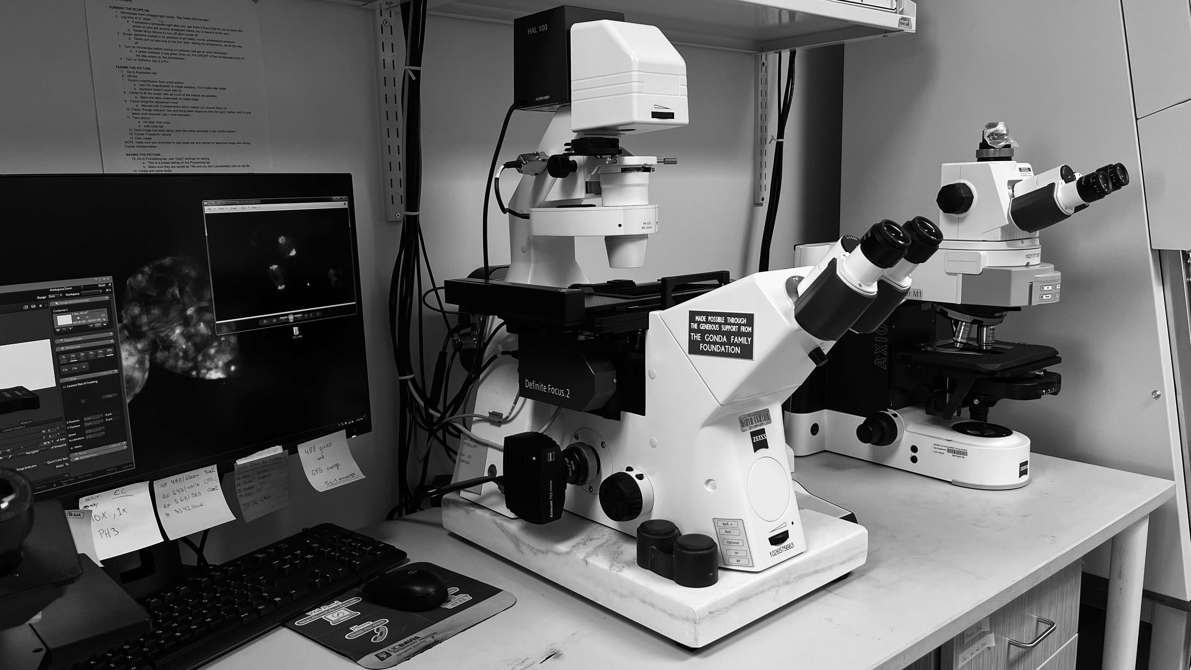
The lab is fully equipped for all aspects of sperm and embryo cell biology and tissue culture. We have a BD AccuriC6 flow cytometer®, with a Minitube SpermVision system for sperm motility analysis. An Olympus BX60 upright fluorescence microscope with Nomarski optics and color digital camera, a Zeiss AxioImager® automated phase contrast microscope with fluorescence is located within the lab.
Two microscope and procedure rooms are located within the lab and houses two Olympus ICSI inverted fluorescence microscopes, Zeiss Zen Blue imaging workstation with Z-focus controllers and 3D software. We have two embryo lab rooms equipped with ICSI workstations fitted with Eppendorf micromanipulators and microinjectors.
The MiriTL® time-lapse imaging embryo incubators and a companion triple gas incubator allow us to monitor growing embryos with minimal interference. An inverted Zeiss Observer fluorescence microscope with live-cell imaging and Zen Blue capabilities is also located within the laboratory.
We also have a Planar computer-controlled programmable cell freezer, a tissue culture laminar flow hood and biosafety cabinet that is also located within the lab, and three CO2 incubators for cell and tissue culture.
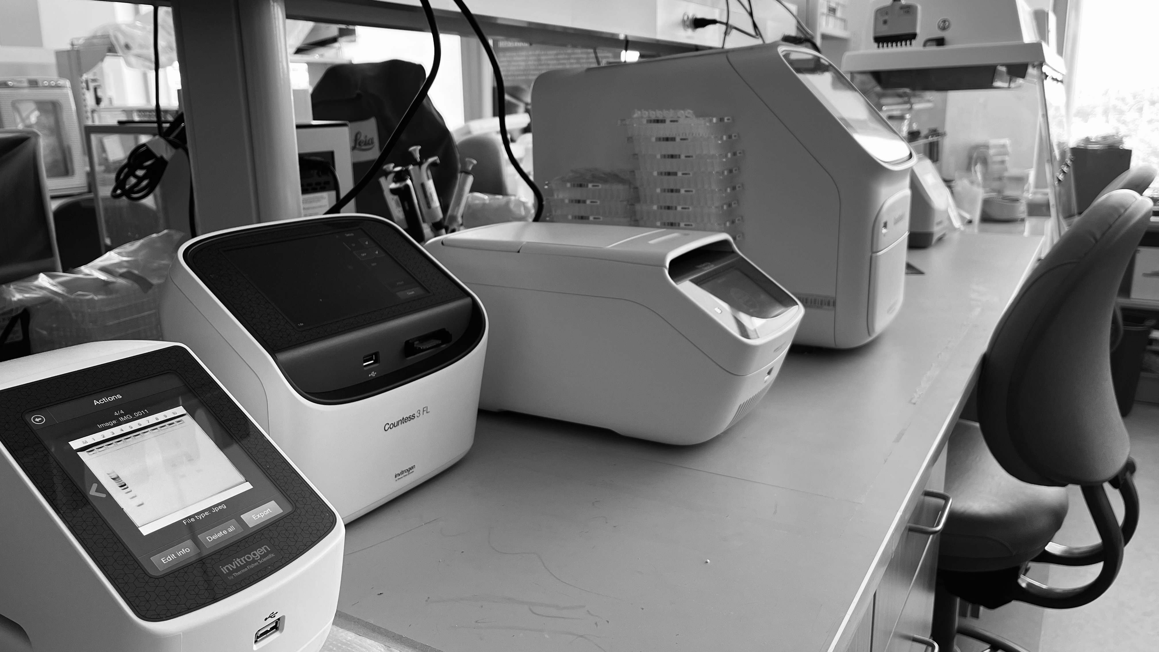
Our laboratory is also equipped with thermocycler, real-time PCR, PCR hood, tissue dissociator (gentleMACS Dissociators), and tissue homogenizer (Bead Ruptor 4).
We also have access to high-performance computing for all our genomic, transcriptomic, and metagenomic analysis.
Click here for map and directions


